Introduction
VWF multimeric analysis is a method for
analysing the concentration and distribution of Von Willebrand Factor [VWF] multimers that are present in plasma. VWF multimer analysis is usually performed after functional and immunological VWF assays indicate an abnormality and rarely if ever in isolation.
VWF is synthesised as a 2050 amino acid monomer [pro-vWF] and the pro-VWF subunits undergo dimersation in the endoplasmic reticulum through disulphide bonding of their C-terminal ends. The VWF dimers then move to the Golgi apparatus where the dimers are assembled by cross-linking of cysteine residues at their N-terminal ends to form a series of VWF multimers of increasing molecular weight.
The largest multimers are found in the in endothelial
cell Weibel–Palade bodies and platelet α-granules [which serve to store VWF prior to its export in the circulation] but are detected only transiently in plasma. Whilst the high molecular weight multimers have the greatest haemostatic capacity, they are also potentially thrombogenic - see ADAMTS13 Assays and TTP.
The catabolism of vWF is mediated primarily by ADAMTS13 - the 13th member of ADintegrin-like And Metalloprotease with ThromboSpondin type 13 motifs - a metalloprotease which limits platelet aggregation and microthrombi formation in the micro-circulation by cleaving Von Willebrand Factor [VWF] between Tyrosine 1605-Methionine 1606 [Tyr1605-Met1606] in the A2 domain, to generate a series of small molecular weight multimers. It is this multimeric pattern that is analysed in the laboratory.
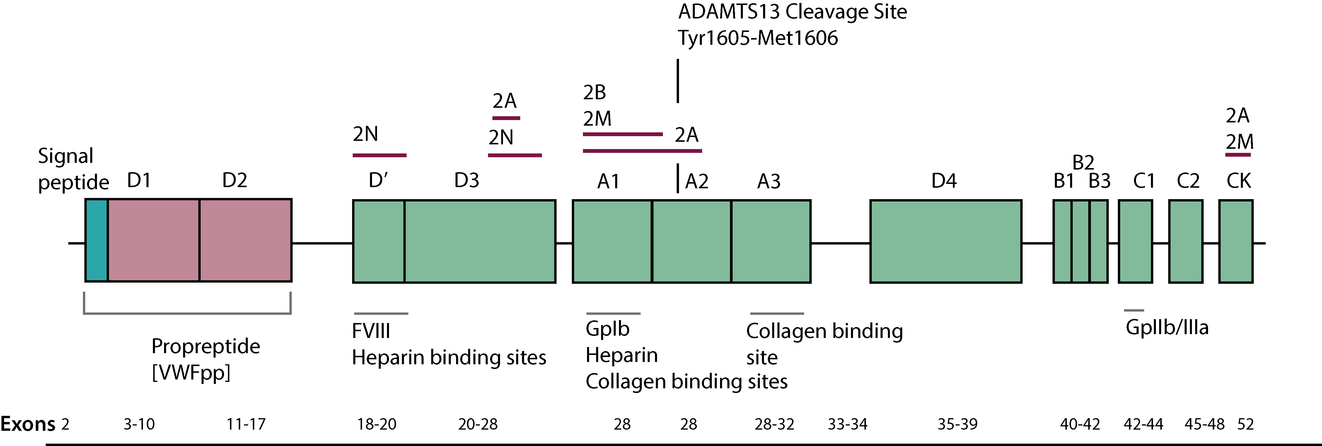
In the absence of ADAMTS13 the cleavage of VWF does not occur and ultra large VWF multimers appear in the plasma. The absence of ADAMTS13 and the generation of ultra large VWF multimers is associated with Thrombotic Thrombocytopaenic Purpura [TTP.]
Principles & Method
1. The first approach to an analysis of VWF multimers involved 2 Dimensional Crossed Immunoelectrophoresis [2D-CIE.]
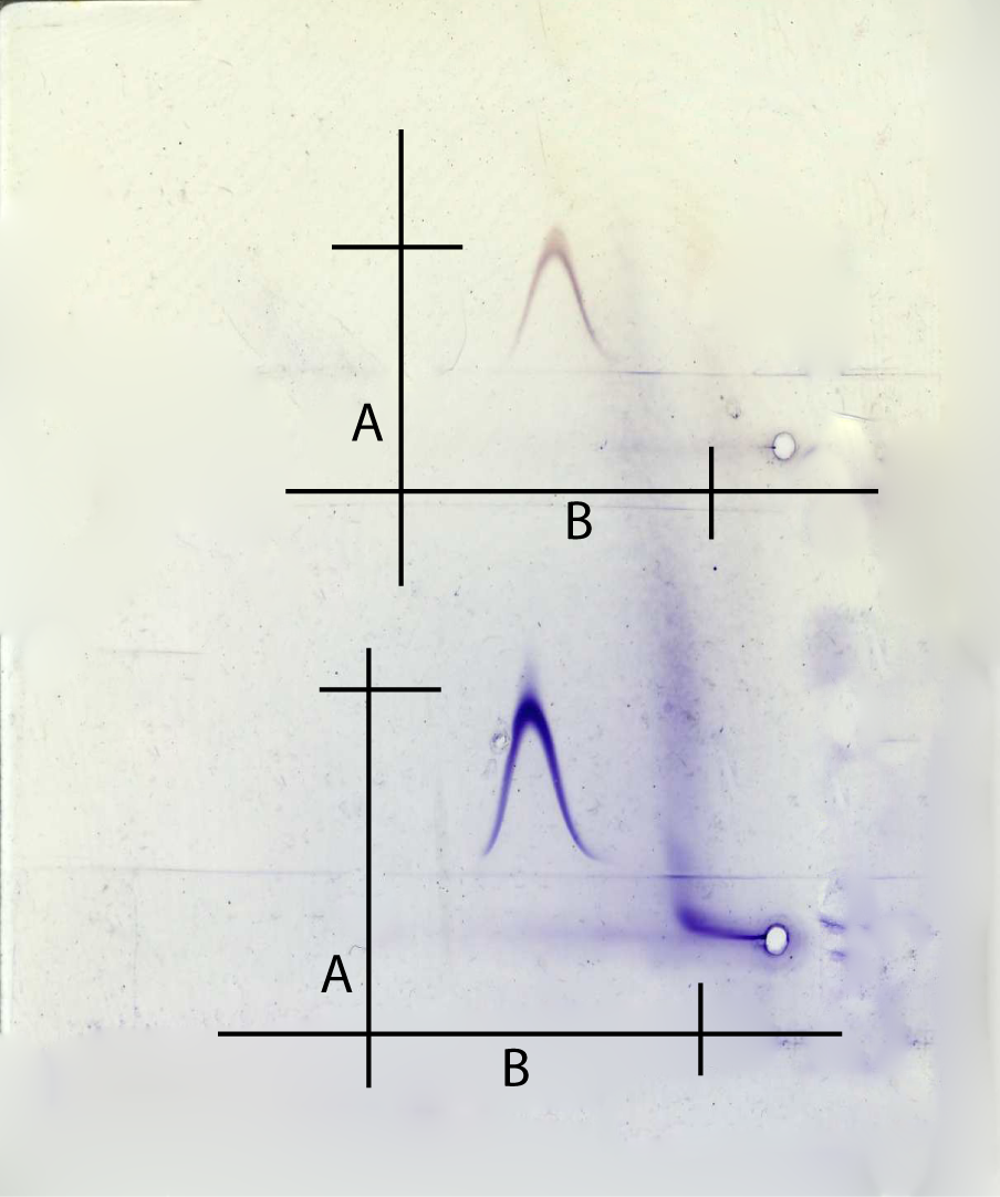
In this gel, VWF has been stained with Coumassie Blue following 2D-CIE. The migration index [MI] can be calculated from the ratio of B:A. In general the MI is <1.25.
2. Multimeric analysis of VWF is carried out by
electrophoresis of plasma samples using non-reducing
agarose gels in the presence of sodium
dodecyl sulphate (SDS). Gels may be of varying concentration e.g. 1.5% for high resolution and 1% of low resolution. Higher gel concentrations
are useful to provide enhanced discrimination of
smaller multimers and individual sub bands. For most diagnostic purposes, low-resolution gels are used.
Following electrophoresis the multimers are visualised:
1. The gels are fixed, washed and dried and then reacted with
an anti-human VWF 125I-labelled
antibody followed by autoradiography.
or
2. Western blotted and then probed with either
enzyme-labelled antibodies and subsequent visualization
of VWF multimers using enzyme substrates
or enhanced chemiluminescence followed by autoradiography.
Interpretation
In normal plasma, VWF multimers appear as a
series of bands separated by the mass of 2 subunits
- see below. Higher resolution gels
reveal the presence of three bands comprising each
multimer (a major band with leading and trailing
sub-bands), the result of proteolytic cleavage in the
circulating blood.
An exception is Vicenza variant
VWD with ultra large VWF multimers and low VWF levels.
VWF Vicenza [also known as Type 1C VWD] arises from increased clearance of the mutant VWF from the plasma.
| VWD Type | Multimer Analysis |
|---|---|
| 1 | All multimers are present including high molecular weight [HMW] forms but all are present in reduced concentration |
| 2A | Absence of large and intermediate forms |
| 2B Platelet-type VWD |
Absence of large forms Platelet-type VWD shows similar results to 2B VWD with a loss of the HMW multimers |
| 2M | All sizes present |
| 2N | All sizes present |
| 3 | Absent multimers as no VWF |
| VWD Vicenza | Ultra-large VWF multimers present |
Reference Ranges
There are no clear reference ranges for VWF multimer analysis as it is primarily a qualitative test. However, densitometric scanning can be performed to quantify the intensity of each multimer and hence the relative concentration of each multimeric band. By comparison against a control it can be seen which multimers are missing or reduced in concentration.
What Test Next
Multimer analysis is performed as part of a series of tests of VWF and rarely in isolation. See Introduction to Von Willebrand Factor for summaries and an algorithm on the investigation of a case of suspected VWD